Symbols:

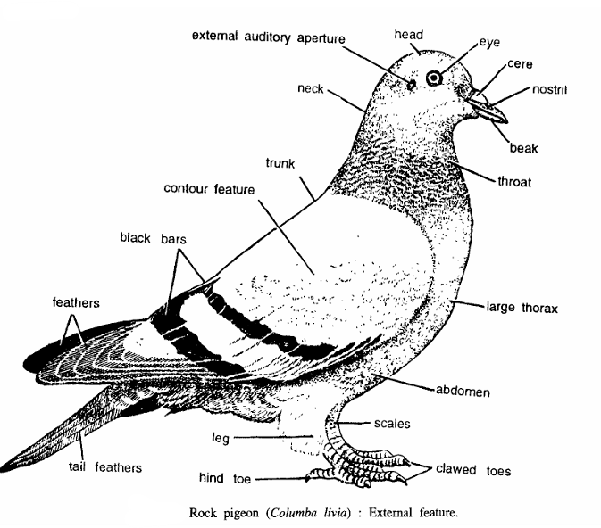
The pigeon is a typical flying bird. It is a preferred laboratory animal for low cost and easy availability.

Killing:

Pigeons are killed with chloroform.

Dissection:

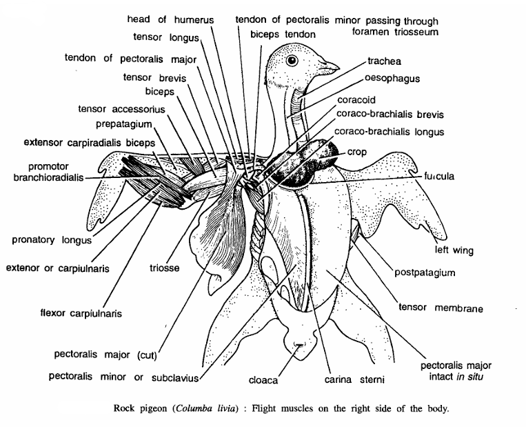
Pluck the feathers and put those in a basket in a corner of the laboratory. Lay the bird on a dissecting tray with the ventral surface up. Fix the pigeon in that position by pushing pins through wings and hind limbs.

Give a longitudinal incision on the skin of the breast along the mid-ventral line.

Continue the incision both anteriorly and posteriorly up to the extremities of the trunk. Give lateral incisions on the skin along the limbs. Separate the skin from the underlying muscles and remove it.

Dissection of Muscles of Flight:

All the flight muscles narrow down anteriorly. While dissecting a muscle, separate it from its attachment but never cut the insertion. The breast of the pigeon is boat- shaped.

The muscles exposed after the removal of the skin are the two pectoralis major, which nearly fill the wedge-shaped space between the body and the keel of the sternum, and form the breast.

With a sharp scalpel cut the attachment of one pectoralis major to the keel, either on the right or left side of it. Posteriorly and laterally the muscle is attached to the body wall by membranous tissue. Cut along the line of at­tachment and turn it upwards.

The pectoralis minor is shorter and nar­rower and dorsal to the pectoralis major. Cut its attachment to the anterior part of the sternum to separate it from the latter. The coracobrachialis or the third pectoralis is the smallest breast muscle for flight. It is attached to the coracoid and the sternum. Sepa­rate it from the coracoid and sternum.

Pectoralis major (depressor muscle):

It is the largest and most ventral flight muscle. The width is maximum at about the middle, where it is thickest also. The fibres of the muscle converge anteriorly to be inserted into the ventral aspect of the humerus, which it depresses.

Pectoralis minor (Supracoracoideus or elevator muscle):

It is wide and thick at the middle. The much narrowed anterior end sends its tendon through the foramen triosseum to be inserted into the dorsal aspect of the humerus, which it elevates.

Coracobrachialis (Scapulo-humeral):

A small, narrow muscle, inserted into the ventral surface of the humerus through its tendon. It helps in depression of the wing.